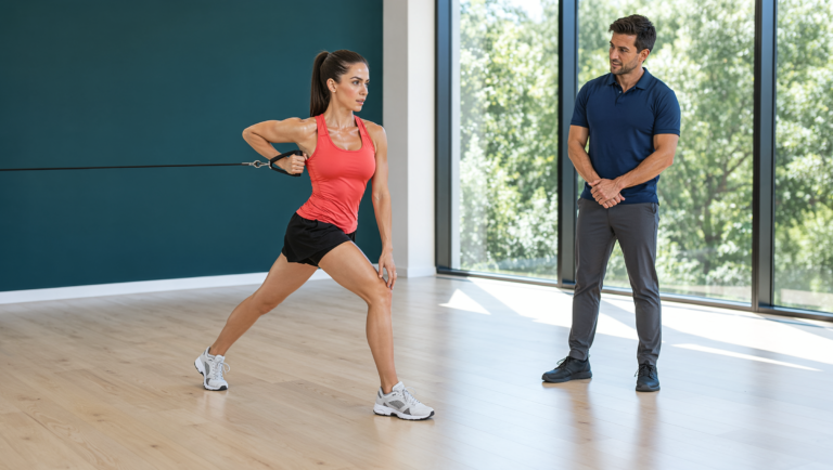
Bone is often thought of as the fixed frame of the body, but it is living tissue that keeps adjusting its mass and architecture to the loads placed on it. That responsiveness is one of the main reasons strength work matters well beyond muscle. The research on exercise and bone mineral density gives a fairly specific picture of which kinds of loading the skeleton responds to, at which sites, and how high-intensity training has been introduced safely in older adults who already have low bone mass.
For nearly 30 years, Temple Human Performance has combined massage and osteopathic-based manual therapy with movement training, physical preparation, and nutrition in one approach to treatment and training. On the personal training side of that work, loading the skeleton is part of the reason we progress strength work the way we do. This article looks at how bone adapts to mechanical load, what pooled trials report about resistance and impact exercise and bone mineral density, what the LIFTMOR trials found in women and men with low bone mass, and how we apply those principles with clients.
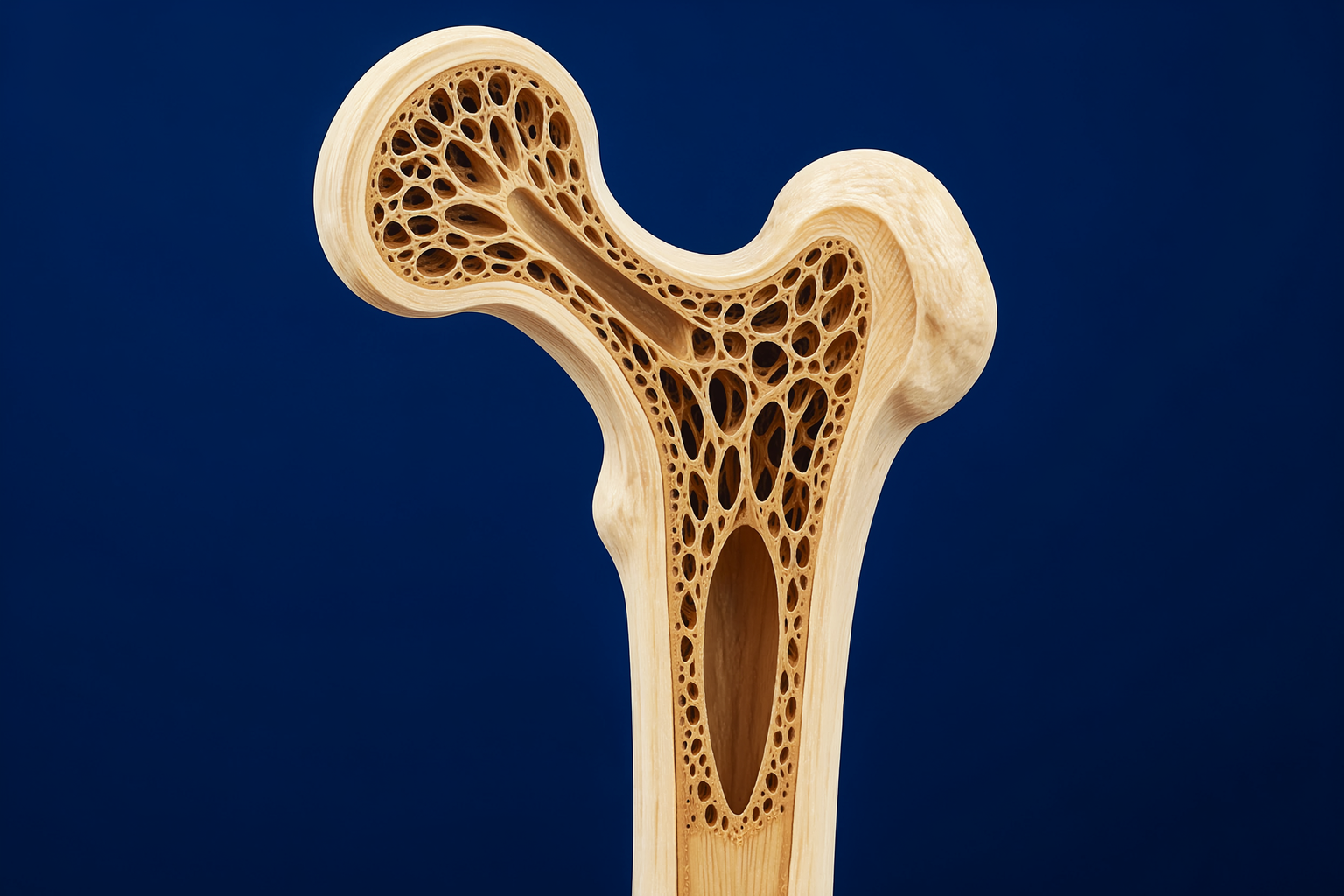
Bone has two main structural forms. Cortical bone is the dense outer shell that gives a bone its strength in bending and compression, and trabecular bone is the lattice of fine struts inside, concentrated in the vertebrae and at the ends of long bones such as the upper femur. Both are continually remodeled, and both respond to the strains that muscle forces and ground contact place on them. In research, bone mineral density (BMD) is usually measured by dual-energy X-ray absorptiometry (DXA) at the lumbar spine and at sites around the hip, such as the femoral neck and the total hip.
Turner (1998) summarized three rules that describe how bone adapts to mechanical stimuli. Adaptation is driven by dynamic rather than static loading. Only a short duration of loading is needed to start an adaptive response. And bone cells accommodate to their customary loading environment, which makes them less responsive to routine signals over time. Taken together, the rules point toward loading that is dynamic, meaningful in magnitude, and different enough from daily routine to register as a new signal, rather than more of the same activity a person already does every day.
The Cochrane review by Howe and colleagues (2011) brought together 43 randomized controlled trials with 4,320 postmenopausal women. For BMD at the femoral neck, the most effective type of exercise appeared to be non-weight-bearing, high-force exercise such as progressive resistance training for the lower limbs. For BMD at the spine, the most effective intervention was a combination program that mixed exercise types. The authors described the overall effect of exercise on bone density as relatively small and statistically significant, but possibly important, and concluded that exercise has the potential to be a safe and effective way to avert bone loss in postmenopausal women.
Earlier meta-analyses by Martyn-St James and Carroll looked at specific forms of loading. Their 2006 analysis of progressive, high-intensity resistance training in postmenopausal women found a significant increase in lumbar spine BMD across 14 trial groups; the femoral neck result was positive but did not reach statistical significance. Their 2009 analysis of impact exercise found that programs mixing jogging with walking and stair climbing, and programs combining impact exercise with high-magnitude resistance exercise, were effective at both the lumbar spine and the femoral neck. Programs that relied on high-impact or odd-impact activity alone were not effective at any site, which is why the authors made the case for mixed loading.
A more recent meta-analysis by Kistler-Fischbacher, Weeks, and Beck (2021) sorted 53 trials, testing 63 interventions in healthy postmenopausal women, by exercise intensity. At the lumbar spine, high-intensity exercise produced a larger BMD effect (a mean difference of about 0.031 g/cm²) than moderate or low intensity (about 0.012 and 0.010 g/cm²). At the femoral neck, low- and moderate-intensity exercise were similarly effective, while no effect was seen for the small number of high-intensity interventions, a result the authors suggested may reflect a lack of statistical power. They concluded that resistance training, potentially combined with impact training, appears to be the most effective bone-building (osteogenic) stimulus at the spine and hip, and that the human data show the same positive relationship between load magnitude and bone response that animal research has long described.
High-intensity loading has traditionally been avoided for people with low bone mass because of a perceived risk of fracture. The LIFTMOR trial, led by Watson and colleagues (2018), tested that assumption directly. It randomized 101 postmenopausal women, average age 65, with low bone mass to eight months of twice-weekly, 30-minute, supervised high-intensity resistance and impact training (HiRIT), performed as five sets of five repetitions at more than 85 percent of one-repetition maximum, or to a home-based, low-intensity exercise program. The HiRIT group’s lumbar spine BMD rose by about 2.9 percent while the comparison group’s fell by about 1.2 percent. Femoral neck BMD rose slightly (about 0.3 percent) in the HiRIT group and fell by about 1.9 percent in the comparison group. HiRIT also produced greater gains in femoral neck cortical thickness, a small difference in height, and better results on every functional measure tested: the timed up-and-go, functional reach, five-times sit-to-stand, and back and leg strength. Compliance averaged 92 percent, and the only adverse event reported in the HiRIT group was a minor lower back spasm. The authors concluded that, under highly supervised conditions, the program enhanced indices of bone strength and functional performance in this group of otherwise healthy women.
A follow-up analysis of the same participants (Watson et al., 2019) examined the spine directly for signs of vertebral fracture. There were no changes in vertebral fracture classification in the HiRIT group after eight months, and standing-tall thoracic kyphosis, the forward rounding of the upper back, decreased by about 6.7 degrees compared with about 1.6 degrees in the comparison group.
The training itself was built on a few fundamental lifts. The published protocol for the men’s version of the trial (Harding et al., 2017) describes sessions built around the deadlift, squat, and overhead press, plus five sets of five jumping chin-ups finished with a firm, flat-footed landing. The same protocol lists the safeguards used in the LIFTMOR trials: full supervision of the high-intensity sessions by a qualified exercise scientist, small group sizes, an initial familiarization period using low-load variations of each exercise focused on lifting technique, and weights that increased progressively with training exposure.
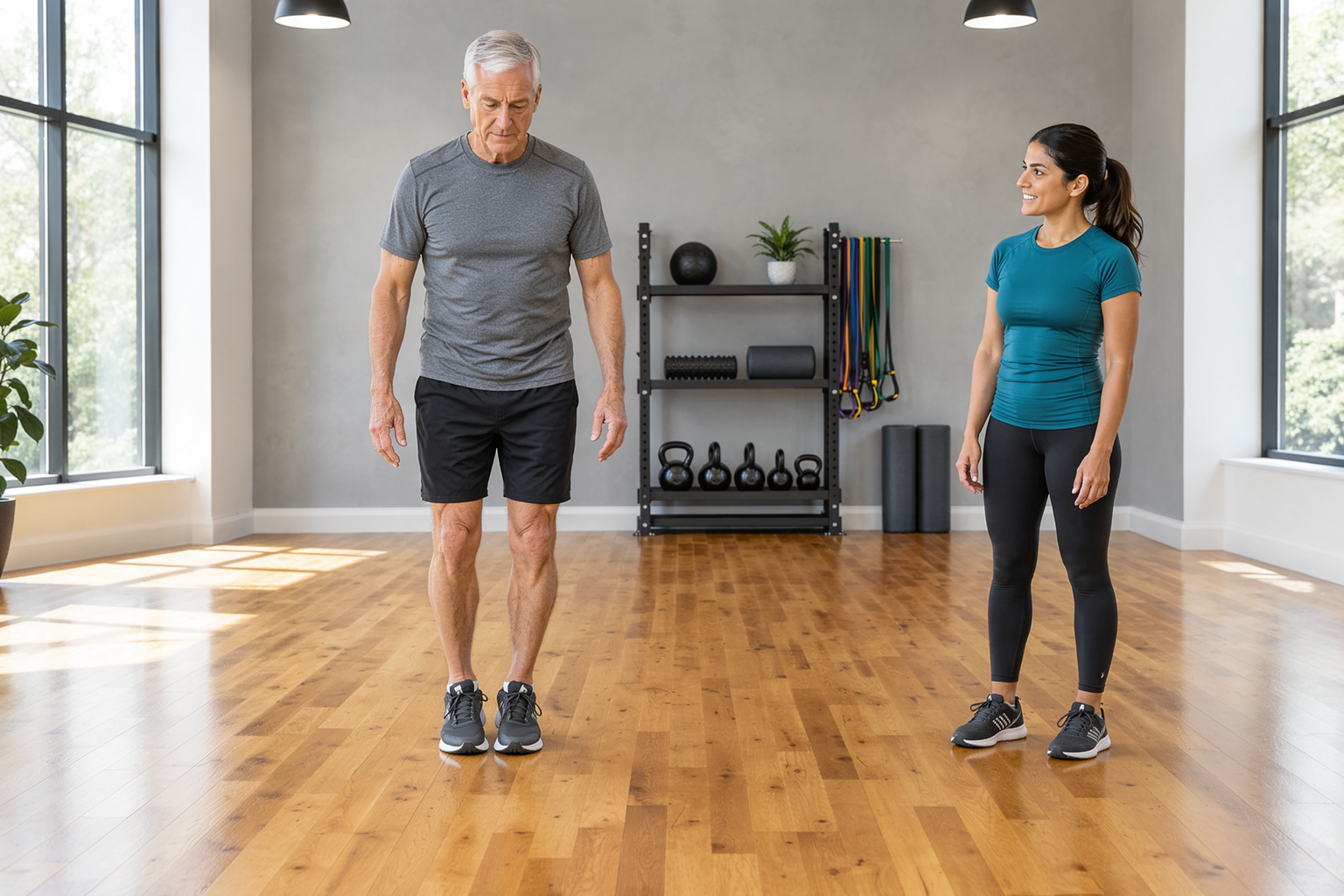
The men’s trial, LIFTMOR-M (Harding et al., 2020), enrolled 93 men, average age 67, with low BMD at the hip or spine. Men were randomized to HiRIT or to machine-based isometric axial compression training and compared with a matched, nonrandomized control group. Compared with the control group, HiRIT improved BMD at the greater trochanter (about 2.8 percent versus about -0.1 percent) and the lumbar spine (about 4.1 percent versus 0.9 percent), along with heel ultrasound measures of bone, lean mass, the timed up-and-go, the five-times sit-to-stand, and back and leg extensor strength. HiRIT also improved lumbar spine BMD more than the isometric program. Compliance was similar in the two training groups, at about 78 percent, and the five adverse events reported across the two programs were minor. The investigators described HiRIT as well tolerated.
The Exercise and Sports Science Australia position statement on exercise for bone health (Beck et al., 2017) draws these findings into practical guidance. It states that evidence from animal and human trials indicates bone responds positively to impact activities and high-intensity progressive resistance training. It also emphasizes that building muscle strength, balance, and mobility reduces the risk of falls, which is particularly relevant for people with limited functional capacity or a very high risk of fracture. The statement recommends that bone-focused programs be accompanied by sufficient calcium and vitamin D, notes that loaded spinal flexion is not recommended, and adds that impact activities may need modification when osteoarthritis or frailty is present. Its central point is individualization: an exercise program for bone should account for existing bone health, other health conditions, and functional and clinical risk factors for falls and fracture.
At Temple, we treat the skeleton as one of the tissues that strength work is meant to reach. The trials point consistently in the same direction: dynamic loading of meaningful magnitude, built around fundamental lifts and, where appropriate, impact, progressed over months rather than weeks. Light, familiar activity has its place, but the research suggests that the loads most likely to register with bone are ones that exceed what a person’s daily routine already provides.
The way the LIFTMOR investigators introduced heavy loading matches how we think about progression. Technique comes first, at loads light enough to learn the pattern well. Load increases as control and confidence grow, and impact is added gradually, starting with simple, controlled landings. Coached sessions give us the chance to watch each repetition closely and adjust the exercise, the range, or the load as needed.
Bone density testing and its interpretation belong with a client’s medical provider. Our role is the training: understanding what a client’s provider has shared, respecting it, and building a plan that fits the individual, as the ESSA guidance recommends. That includes the positional details that matter for the spine, such as lifting with a neutral trunk rather than under loaded spinal flexion, and modifying impact when the joints call for it.
The hands-on work has a place in this as well. When a stiff hip, ankle, or upper back limits the positions that squatting, hinging, or pressing require, table work and joint mobilization can open movement options so the loading that follows is well organized. Nutrition coaching supports the same goal from another side, since the guidance pairs bone-focused training with adequate calcium and vitamin D.
Physiokinetix, the education and movement system I developed, views movement as the product of many interacting factors, including tissue loading, motor control, strength and endurance, coordination, and the demands of the task and the environment. Bone belongs in that picture as a tissue whose capacity follows the loads it regularly meets. The research on bone adaptation is a clear example of a principle that runs through all of our work: capacity is built by appropriate, progressive demand.
The PKT framework progresses through Mobility, Stability, Power, and Performance. Mobility opens access to the positions that loaded movement requires. Stability builds the control and balance that make heavier work safe and that the ESSA guidance connects to a lower risk of falls. Power introduces greater force production and progressive loading, which is where the bone-loading findings apply most directly. Performance carries that capacity into daily life, recreation, work, and healthy aging. The levels build on one another, but they are not a rigid protocol, and a client may work in more than one level at a time.
Physiokinetix also holds to the principle of knowledge, not diagnosis, and it links professional sessions with self-care so that clients take an active part between visits. In this context that means explaining what the research shows about bone and load, teaching the lifts and landings well, and helping clients carry that practice into their PKT Self-Care work, so the skeleton keeps receiving a meaningful signal week after week.
Bone adapts to the loads placed on it, responding best to dynamic loading that is meaningful in magnitude and different from routine activity. Pooled trials in postmenopausal women show small but potentially important effects of exercise on bone mineral density, with progressive resistance training and mixed programs that combine resistance and impact performing best, and higher-intensity exercise showing the largest effect at the lumbar spine. The LIFTMOR trials found that supervised, high-intensity resistance and impact training, introduced through careful technique work and progressive loading, improved bone mineral density, indices of bone strength, and functional performance in older women and men with low bone mass, with few and minor adverse events. Exercise guidance adds the importance of strength and balance for fall risk, adequate calcium and vitamin D, sensible spinal positioning, and individualization. At Temple, we build those principles into how strength work is introduced and progressed, how the hands-on work supports the positions that loading requires, and how clients continue the practice on their own, so treatment and training reach bone as well as muscle.