

Clinical context
Strength often arrives before the tendon is ready for it. In the first weeks of a new training block, a client may lift more, jump higher, or hold a heavier position than a month earlier, while the tendons that carry that force are still adapting at a slower pace. This gap between how quickly muscle and tendon respond to training is one of the more practical ideas in loading science, and it shapes how we plan progression for healthy, active people.
At Temple Human Performance, we combine massage and osteopathic-based manual therapy with movement training and physical preparation in one integrated approach. Tendon adaptation sits at the meeting point of those pieces. Hands-on work and active movement help a client move and feel well enough to train, and the training itself is what changes the tendon. How that training is dosed and sequenced determines whether muscle and tendon develop together.
Scientific background
A tendon connects muscle to bone and transmits the force the muscle produces. Its stiffness describes how much it stretches under a given force. A stiffer tendon transmits force more directly, while a more compliant tendon stretches more under the same load. Strain is the amount a tendon lengthens relative to its resting length, and it is a useful way to describe how much mechanical demand the tendon is experiencing.
Stiffness can change in two ways. The tendon can become larger, increasing its cross-sectional area, or the tissue itself can change its material properties, described by Young’s modulus. A tendon with a higher modulus is stiffer per unit of size, much like a rope woven from stronger fibers rather than simply a thicker rope.
Muscle and tendon are built to work as one unit, but they do not adapt on the same timeline or to exactly the same stimuli. Muscle can gain strength quickly, partly through changes in how the nervous system recruits it and partly through growth. Tendon tissue remodels more slowly and responds most to the magnitude of mechanical strain it experiences. When strength rises faster than tendon stiffness, the tendon stretches further during a maximal effort. That is the muscle-tendon mismatch.
What the research shows
Two meta-analyses of healthy adults set the foundation. Bohm and colleagues (2015) pooled 27 studies with 37 training interventions on the Achilles or patellar tendon in 264 healthy adults aged 18 to 50. Training increased tendon stiffness (standardized mean difference 0.70), Young’s modulus (0.69), and cross-sectional area (0.24). Stiffness adaptation depended on loading intensity but not on contraction type, and effects were descriptively larger in programs lasting 12 weeks or more.
Lazarczuk and colleagues (2022) updated this picture with 61 studies and 763 participants, most using resistance training. Loading produced moderate increases in stiffness (0.74), large increases in modulus (0.82), and small increases in cross-sectional area (0.22). Meta-regression showed that modulus, not size, was the main driver of increased stiffness. Higher-strain resistance protocols produced greater increases in modulus and stiffness than lower-strain protocols, and the differences were greater in adult participants than in other age groups. Together, these reviews indicate that healthy tendons are responsive to training, and that most of their early change happens inside the tissue rather than in its size.
The mismatch itself has been studied directly. Lambrianides and colleagues (2024) had 12 young adults train the calf muscles of each leg for 12 weeks with repeated isometric plantarflexion contractions at 80 percent of maximum until failure. One leg trained with the ankle in a plantarflexed position, where the muscle-tendon unit is short and Achilles tendon strain is lower. The other trained with the ankle dorsiflexed, where the unit is long and tendon strain is higher. Muscle adapted similarly in both legs. The tendon, however, increased in stiffness, modulus, and size only in the long-length, higher-strain condition. In the short-length leg, maximal Achilles tendon strain rose steadily over the training period because the muscle grew stronger while the tendon did not change. The authors concluded that muscle adapts faster than tendon and does not need high mechanical load to do so, while tendon depends on strain magnitude.
Mersmann, Bohm, and Arampatzis (2017) reviewed the broader evidence and reached a similar conclusion. Muscle and tendon differ in both the time course of adaptation and their responsiveness to particular stimuli. Plyometric loading, for example, appears to be a stronger stimulus for muscle than for tendon. The authors described a specific tendon loading concept: five sets of four contractions at 85 to 90 percent of maximal isometric effort, with each contraction providing about 3 seconds of high tendon strain.
That concept has been tested in young athletes, the group in which the mismatch has been studied most closely. Domroes and colleagues (2023) followed adolescent athletes across a competitive season and found that around 40 percent of both female and male athletes showed high levels of patellar tendon strain, above 9 percent, during maximal efforts, indicating imbalances between muscle strength and tendon stiffness in both sexes. In a follow-up trial, Domroes and colleagues (2024) added personalized tendon exercises three times per week for about 31 weeks to the training of male adolescent elite athletes, with intensity set to produce about 6.2 percent tendon strain. Compared with athletes who continued their usual training, the intervention group increased both knee extensor strength and normalized patellar tendon stiffness and showed smaller fluctuations in maximal tendon strain across the season.
Age changes the details, though not the principle. Létocart and colleagues (2024) trained 27 healthy men around 70 years old for 12 weeks with either moderate loads at 55 percent of one-repetition maximum or loads progressing from 55 to 80 percent, with volume matched. Both groups increased Achilles and patellar tendon size and tendon force, and stiffness and modulus increased in the Achilles tendon and the deep aponeurosis of the vastus lateralis. The two loading strategies produced similar results.
These studies have limits. Most are small, many measure the Achilles or patellar tendon only, and tendon properties are estimated with ultrasound and dynamometry, which vary between laboratories. The youth studies describe imbalances and their reduction, while the link between imbalance and later tendon problems remains a hypothesis the authors raise rather than an established outcome. The overall direction is consistent: muscle adapts quickly and broadly, tendon adapts more slowly and mainly to sufficient strain, and training can be organized to keep the two in step.
Functional significance
Three points carry the most weight for planning training.
Strength gains can outpace tendon readiness. The early weeks of a new program, a return to sport after time off, or a jump in plyometric volume are periods when muscle may be ahead of tendon. Progression during these windows benefits from patience, especially in high-speed and jumping work that loads tendons heavily.
Heavy, sustained loading targets the tendon. Across reviews, the strain magnitude and intensity of loading mattered more than whether a contraction was isometric, concentric, or eccentric. Longer-length positions and sustained high-effort contractions appear to give tendon tissue the signal it responds to, while lighter, fatiguing work builds muscle without the same tendon response.
Time is part of the dose. Tendon changes in these studies were measured over 12 weeks or more, and the personalized youth program ran for most of a season. Tendon work is most useful as a consistent thread in training rather than a short phase.
Application at Temple Human Performance
When we plan a client’s progression, we consider tendon timelines alongside strength. A client whose strength rises quickly in the first weeks of a program, or who is returning to running, jumping, or court sport, is someone for whom we hold back the rate at which speed and impact increase while the tendon work catches up.
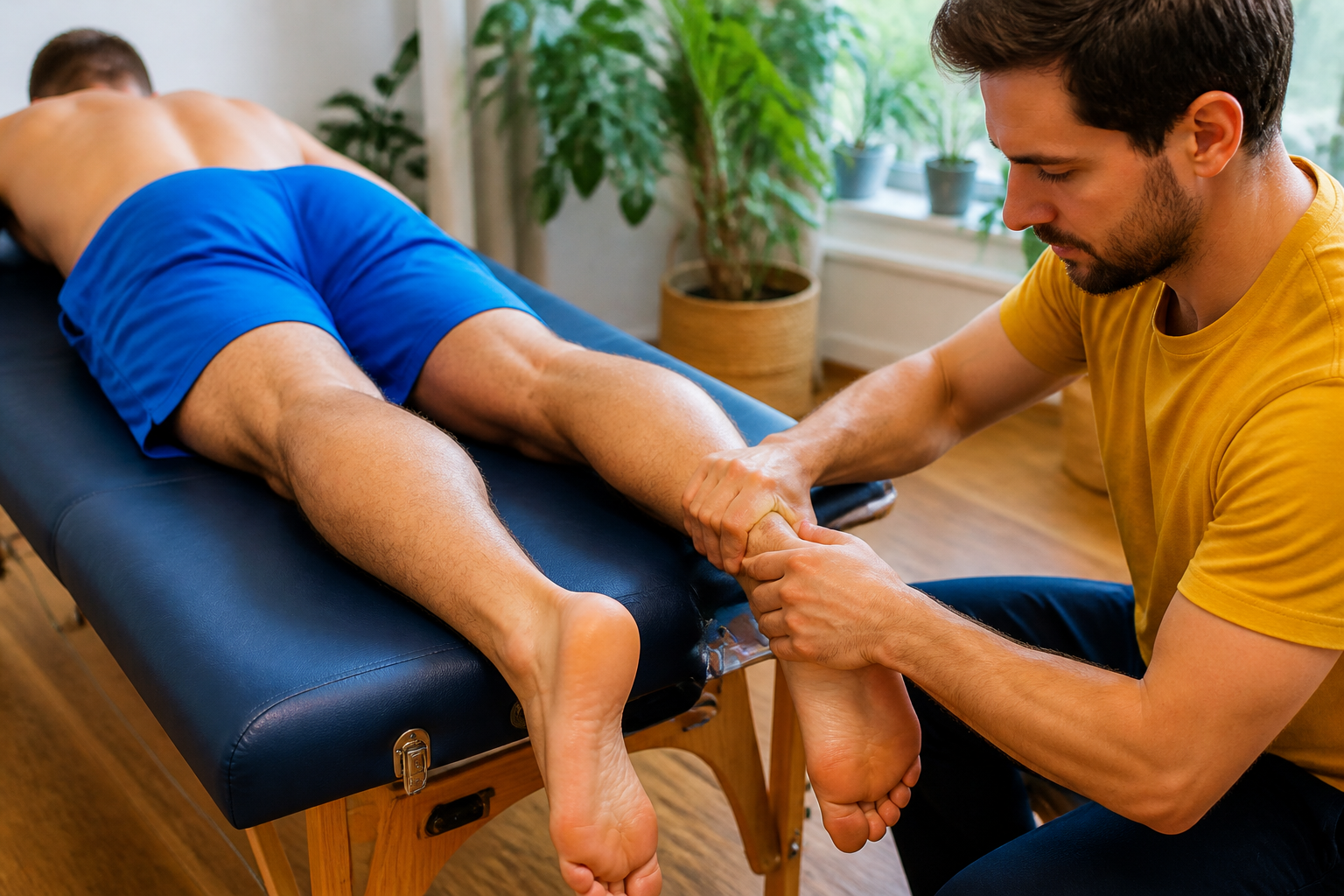
The table work supports that process. Hands-on work around the calf, the front of the thigh, and the surrounding tissue can improve comfort and range, and we follow it with active movement so the change is used rather than left on the table. Ankle and knee range matter here, because the long-length positions that load tendon well depend on reaching those positions comfortably.
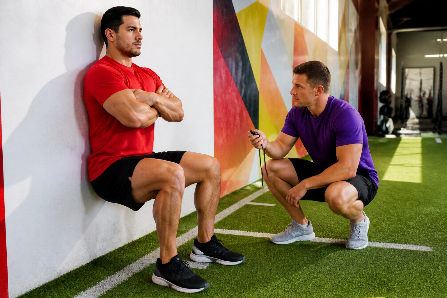
Loading for tendon is deliberate and heavy relative to the individual. Sustained isometric holds in longer muscle-tendon positions, slow heavy calf raises from a deficit, split squat and wall-sit holds for the patellar tendon, and slow resistance work give the tendon a strong strain signal. For heavier holds where a client anchors or pulls against resistance, the PKT Xtreme resistance bands offer a portable way to create high, steady tension. We pair this with the client’s other training and adjust intensity to the individual, since older adults in the research adapted well to moderate, slow loading.
As tendon capacity develops, we build back toward speed: faster lifts, hopping, bounding, landing, and sport-specific movement. Plyometric volume increases gradually because it challenges tendon more than it builds it, and because muscle tends to keep pace with those demands sooner than tendon does.
The Physiokinetix perspective
Physiokinetix organizes movement development through four progressive levels: Mobility, Stability, Power, and Performance. Mobility gives the body access to range, including the longer ankle and knee positions where tendon is loaded well. Stability develops control in those positions so a client can hold and manage high effort safely. Power introduces greater force, resistance, and load tolerance, and this is where the bulk of heavy tendon loading lives. Performance applies those capacities to running, jumping, sport, work, and daily life. The levels build on one another, but they are not a rigid protocol, and tendon timelines are one reason a client may stay longer at one level before moving to the next.
Progressive loading within PKT emphasizes appropriate exposure to physical demand rather than avoiding load, and the variables of progression include resistance, duration, range, speed, and complexity. The muscle-tendon research adds a specific reason to manage those variables carefully: different tissues in the same chain change at different rates. Within the PKT architecture of Assess, Influence, Activate, Load, Integrate, and Perform, this sits mainly in the Load and Integrate stages, where the goal is lasting capacity rather than a short-term gain in strength alone.
The PKT Self-Care program extends this between sessions. Simple tools such as heavy bands, straps, and anchors allow clients to perform sustained, high-effort holds and slow loading at home, keeping tendon work consistent across the weeks it takes to adapt.
Conclusion
Healthy tendons adapt to training, and most of that adaptation reflects changes in the tissue’s material properties rather than its size. Tendon responds mainly to the magnitude of strain it experiences, while muscle gains strength faster and under a wider range of stimuli. When strength rises ahead of tendon stiffness, the tendon stretches further during maximal effort, and research in young athletes shows that targeted, high-strain tendon loading can keep the two in better balance. Within our approach, we use hands-on work and active movement to open access to useful positions, apply heavy and sustained loading to build tendon capacity, progress speed and impact at a pace the tendon can match, and give that work enough time to take hold.
References
- Bohm S, Mersmann F, Arampatzis A. Human tendon adaptation in response to mechanical loading: a systematic review and meta-analysis of exercise intervention studies on healthy adults. Sports Med Open. 2015;1(1):7. https://doi.org/10.1186/s40798-015-0009-9
- Lazarczuk SL, Maniar N, Opar DA, Duhig SJ, Shield A, Barrett RS, Bourne MN. Mechanical, material and morphological adaptations of healthy lower limb tendons to mechanical loading: a systematic review and meta-analysis. Sports Med. 2022;52(10):2405-2429. https://doi.org/10.1007/s40279-022-01695-y
- Lambrianides Y, Epro G, Arampatzis A, Karamanidis K. Evidence of different sensitivity of muscle and tendon to mechano-metabolic stimuli. Scand J Med Sci Sports. 2024;34(5):e14638. https://doi.org/10.1111/sms.14638
- Mersmann F, Bohm S, Arampatzis A. Imbalances in the development of muscle and tendon as risk factor for tendinopathies in youth athletes: a review of current evidence and concepts of prevention. Front Physiol. 2017;8:987. https://doi.org/10.3389/fphys.2017.00987
- Domroes T, Weidlich K, Bohm S, Arampatzis A, Mersmann F. Effect of sex on muscle-tendon imbalances and tendon micromorphology in adolescent athletes: a longitudinal consideration. Scand J Med Sci Sports. 2023;33(12):2561-2572. https://doi.org/10.1111/sms.14483
- Domroes T, Weidlich K, Bohm S, Mersmann F, Arampatzis A. Personalized tendon loading reduces muscle-tendon imbalances in male adolescent elite athletes. Scand J Med Sci Sports. 2024;34(1):e14555. https://doi.org/10.1111/sms.14555
- Létocart AJ, Couppé C, Mabesoone F, Charleux F, Marin F, Dermigny Q, Magnusson SP, Svensson RB, Grosset JF. Effects of moderate and heavy slow resistance training on Achilles and patellar tendons and muscles aponeuroses in elderly men. Scand J Med Sci Sports. 2024;34(11):e14766. https://doi.org/10.1111/sms.14766